CLASS II FOIL - UPPER FIRST BICUSPID -
MESIAL
Class II cavity prepared in
upper first bicuspid, mesio-occlusal surfaces, for
the reception of gold foil as the restorative medium.
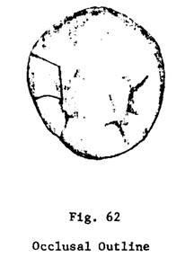
Outline Form
A. Occlusal
surface (Fig. 25)
1.
Structural defects and any undermined enamel are removed. Entire central groove
area is included.
2.
Surrounding walls are extended to provide for the manipulation of the gold foil
and the proper finishing of the margins of the restoration.
3.
Distal wall parallels the distal marginal ridge and meets the buccal and lingual walls at a definite sharp angle.
4. Buccal and lingual walls extend to blend harmoniously with
the corresponding walls of the proximal portion of the preparation, and to
provide for access to the interior of the proximal portion of the cavity during
insertion of the gold foil. This usually requires a reverse curve in the buccal wall where the occlusal
portion meets the proximal wall.
5. The average bucco-lingual width of the isthmus of this preparation is
no greater than 3/4 to 1 mm in natural tooth. (The isthmus is the narrowest bucco-lingual dimension of the step portion.)
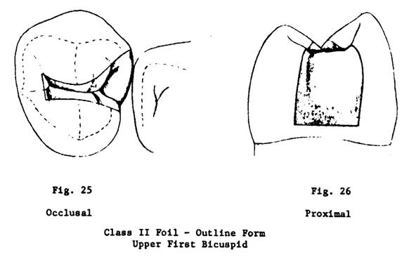
B. Proximal surface (Fig. 26)
1. Gingival Wall
a.
Placed so gingival margin is uniformly about halfway into the depth of the free
gingival crevice.
b.
Straight bucco-lingually.
c. Meets
the buccal and lingual walls at a definite angle. The
bucco-gingival angle is usually approximately a right
angle or obtuse angle. The linguo-gingival angle is
acute.
Note: In the development of the outline form it is essential that the
gingival wall be established first. The position occupied by the gingival wall
determines the outline form of the entire preparation. This maxim holds true
for all proximal cavity preparations.
2. Buccal
and Lingual Walls
a.
Include defective or carious area.
b.
Extended to areas of the tooth surface which are less susceptible to recurrence
of decay.
c. Placed
in areas which are capable of being finished readily.
d.
In view of the demands of esthetics and inconspicuousness, in the natural tooth
the buccal margin clears the approximating tooth by
1/2 - 3/4 mm. Viewing it at an angle perpendicular to the tangent it is
straight from occlusal to gingival; sighting it from
over the mesial approximating tooth it is, in
general, parallel to the contour of that tooth.
e.
The lingual margin uniformly clears the approximating tooth by 1 mm.
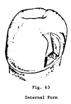
Resistance and Retention Form
A. Occlusal
(step) portion
1. Pulpal Wall
a. Flat.
b. At
right angles to the long axis of the tooth (Fig. 28).
c. Established just within the dentin (Fig. 27).
2. Buccal and Lingual Walls
a. Smooth and straight.
b. Meet the pulpal wall at a
right angle except at the isthmus where the angle is slightly obtuse (Fig. 28).
3. Buccal wall follows into disto-buccal
developmental groove slightly, to form a dovetail (Fig. 25).
4.
Distal Wall
a.
Straight.
b. Parallels the distal marginal ridge (Fig. 25).
c. Meets the buccal and
lingual walls at sharp angles.
d. Forms an obtuse angle with the pulpal
(Fig. 27).
5.
The reverse curve in the buccal outline follows
through the whole wall as the occlusal portion meets
the proximal portion. This opens the "throat" of the cavity and
allows better access with the condensing instruments (Fig. 28).
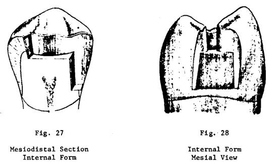
B. Proximal portion (Fig. 28)
1.
Gingival Wall
a. Flat, straight.
b. Basically at right angles to the long axis of the
tooth bucco-lingually, dependent on requirement that
this margin must be under the normal free gingival tissue.
c. Slopes in to the axial to meet that wall at a
slightly acute angle (Fig. 27).
d. Axio-gingival line angle
is established just within the dentin but is slightly deeper axially than in a
preparation for a gold inlay.
e. Meets the buccal and
lingual walls in clean sharp definite angles.
2. Buccal and Lingual Walls
a. Meet the axial wall at a right angle or very
slightly obtuse angle (Fig. 28).
b. Are on a plane parallel with that of the enamel
rods.
3.
Axial Wall (Fig. 27)
a.
Parallel with long axis of tooth (not with surface contour as in the Class V
Foil preparation).
b. Flat bucco-lingually.
c. Established within dentin.
d. Meets pulpal wall at
definite sharp angle.
4. Bucco-axio-gingival and linguo-axio-gingival
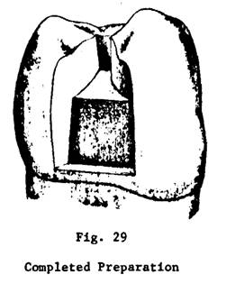
point angles are definite, sharp and slightly acute (Figs. 27 & 29).
5.
Bucco-axial line angle and linguo-axial
line angle fade into buccal and lingual walls
respectively of the occlusal portion of the cavity
(Fig. 28).
C. Retention, then, is
provided by
1.
Convergence occlusally of buccal
and lingual proximal walls.
2. Occlusal dovetail.
3.
Slightly acute axio-gingival line angle.
4.
Compressibility of dentin.
Convenience Form
Is
necessary to enable adequate line of force for compaction of the gold foil. The only special provision is the establishment of
the reverse curve in the buccal wall. Otherwise the
requirements are met by having clean sharp line angles, point angles and proper
outline form.
Finish of the Enamel Walls
1. The enamel walls are
planed smooth.
2. Walls must have full
length enamel rods, supported by sound dentin.
3. A very light bevel is
usually required on the occlusal cavosurface
margin, and is sometimes needed on the gingival cavosurface
margin to avoid short unsupported enamel rods (Figs. 27, 29).
4. The cavosurface
angle presents a continuous, smooth line devoid of irregularities.